산전 초음파 검사와 태아 신우확장, Prenatal ultrasonography and renal pelvis dilatation in the fetus
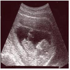
사진 1-130. 자궁 속에서 자라고 있는 임신 2개월 된 태아의 초음파. 테스트 튜브에서 수정된 후 이제는 엄마의 자궁 속에서 잘 자라고 있다.
Copyright ⓒ 2011 John Sangwon Lee, MD., FAAP
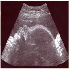
사진 1-131. 자궁 속에서 자라고 있는 임신 7개월 태아의 초음파. 테스트 튜브에서 수정된 후 이제는 엄마의 자궁 속에서 잘 자라고 있다. 눈, 얼굴, 입 모양도 볼 수 있다.
Copyright ⓒ 2011 John Sangwon Lee, MD., FAAP
- 미국에서는, 임산부들의 75%가 임신 중 적어도 한번 내지 한번 이상 산전 초음파 검사를 받는다.
- 의술과 과학 기술의 발달, 임신과 태아의 이상의 유무 진단, 임산부의 요구 등으로 산전 초음파 검사를 많이 한다.
- 임신 중 양수검사 대신 산전 초음파 검사를 요즘 더 많이 한다.
- 산전 초음파 검사로
-
- 태아의 선천성 기형이나 태아에게 생겨 있는 질병을 출생 전 진단할 수 있고
- 산과 전문의와 주산기과 전문의, 소아청소년과 전문의는 그런 선천성 기형이나 병을 가지고 아기가 태어나기 전에 태아 응급 의료 치료팀의 파트너가 되어 태아의 건강문제를 보다 더 효과적으로 다룰 수 있다. 또 출생 당시 신생아의 건강 문제를 즉시 적절히 진단 치료할 수 있다.
- 태아에게 선천성 기형이나 질병 등이 산전 초음파 검사에서 발견되면 그 태아가 출생하기 바로 전 신생아 전문의와 신생아 전문적 심폐소생술 팀 등이 갓 태어난 신생아에게 있을 수 있는 건강문제를 즉시 도와줄 준비를 미리 할 수 있다.
- 또 성장지연이 자궁 내 태아에게 발견되면 그 원인을 알아 그 원인에 따라 적절히 치료 하고 분만 전 임산부는 태아 건강관리에 더 조심하고, 산전 임신 건강 관리를 더 잘하고, 또 출생하기 전까지 태아를 더 특별히 조심히 관찰한다.
- 산전 초음파 검사의 결과가 경도 비정상으로 나타나는 경우 때로는 출생 후 아무 이상이 발견되지 않는 경우도 있다.
- 그 때문에 산전 초음파 검사에 관해 더 많은 연구가 요한다고 한다.
- 이런 이유로 태아 초음파 검사의 결과가 경도로 비정상이지만 확실히 모를 때는 아기가 태어나기 전 소아청소년과 전문의, 주산기과 전문의와 산부인과 전문의가 의료팀의 파트너로 협력해서 진단 치료하는데 문제가 있다.
- 또 산전 초음파 검사의 결과에 따라 임산부를 카운슬링 하는 데 문제가 생길 수 있다
- 태아의 신우확장 등 태아 선천성 기형을 진단 평가하는데 산전 초음파 검사는 대단히 가치가 있다.
- 다운 증후군 및 염색체 이상으로 생긴 다른 선천성 기형, 신우나 방광, 또는 요도 출구의 폐쇄로 인한 방광 요관 역류나 수신증 등을 진단하는 데 산전 초음파 검사는 중요한 역할을 한다.
- 산전 초음파 검사에 나타난 대뇌 뇌실 확장, 반향적 위장관, 반향적 심장 부위, 양수 결핍증, 융모막 낭종 등은 임상적으로 별 의미가 없는 경우가 많다.
- 산전 초음파 검사로 쉽게 확실히 진단할 수 있는 태아 신우확장에 대해서 다음에 더 설명한다.
| 태아 신우확장 |
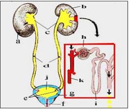
그림 1-132. 비뇨계 해부도
a-우 신장, b-좌 신장, c-신우, d-요관, e-요관구, f-요도, g-방광, h-사구체, i-헨레계제, j-소변, k-혈관
Copyright ⓒ 2011 John Sangwon Lee, MD., FAAP
- 태아 신우확장은 산전 초음파 검사에서 태아들의 2-7%에서 발견된다.
- 그 중 상당수는 출생 전 자연히 없어질 수 있다.
- 신우나 방광, 또는 요도 출구가 선천적으로 막힐 때 방광 요관 역류나 신우확장이 생길 수 있다.
- 임신 제 3기에 산전 초음파 검사를 반복할 때 신우 확장이 없어졌는지 확인하고
- 출생 바로 전 산전 초음파 검사를 추적 재 반복 검사 했을 때 신우확장이 자연히 없어져서 더 이상 나타나지 않으면 생후 다른 검사를 더 이상 할 필요가 없고 관찰적 치료만 해도 된다고 한다.
- 초음파 검사 상 신우확장이 계속 나타나면 출생 후 다운 증후군 등 염색체 이상이 있나 자세히 진찰해야 한다.
- 다운증후군을 가진 신생아에서 신우확장이 35% 발견된다.
- 생후 3일 이후 신생아의 신장, 신우, 요관, 방광 초음파 검사를 하고 출생 후 초음파 검사의 결과가 정상이라도 방광, 요관 역류 등 비뇨기계에 선천성 기형이 있나 알아보기 위해 배뇨 방광 요도 조영술(VCUG) 검사를 출생 후 하는 것을 권장한다.
- VCUG검사로 방광 요관 역류를 진단하기 전에 적절한 항생제로 요로 감염을 예방하는 것이 보통이다.
| 다음과 이유로 산전 초음파 검사를 할 수 있다. |
1. 태아의 생존 능력, 태아의 건강상태와 성장 정도를 알기 위해
- 제왕절개 수술분만,
- 양수검사,
- 유도분만 등을 하기 위한 임신 일수를 알기 위해서,
- 임신자궁의 크기와 임신 일수의 차이가 있을 때 정확히 알기 위해,
- 태아의 선진부를 알기 위해,
- 양수의 양을 알기 위해,
- 염색체 이상이나 태아의 선천성 기형의 여부,
- 임신 40주 이후 태아 건강상태 등을 알아보기 위해서 산전 초음파 검사를 할 수 있다.
2. 태아가 건강에 해로운 인자에 노출됐을 때
- 자궁 외 임신이나
- 포충상몰이 의심될 때,
- 전치태반이나 태반조기 박리,
- 조기 양수파열이나
- 조기 분만,
- 다태 임신,
- 고령 임신,
- 임신성 당뇨병,
- 고혈압,
- 혈관계 질병,
- 산욕증,
- 골반 내 덩어리,
- 자궁의 이상,
- 자궁 섬유종,
- 태아기형을 일으킬 수 있는 알코올, 마약, 유해성 물질에 태아가 노출되었을 때,
- 임신 중 감염병을 알았을 때,
- 태아 적아구증이 있을 때 산전 초음파 검사를 할 수 있다.
3. 그 외 이유
- 원인을 모르는 자궁출혈
- 자궁 통증
- 태동의 감소
- 비정상 생화학적 검사의 결과가 양성일 때 산전 초음파 검사를 할 수 있다.
Prenatal ultrasonography and renal pelvis dilatation in the fetus 산전 초음파 검사와 태아 신우확장
Photo 1-130. Ultrasound of a 2-month-old fetus growing in the womb. After fertilization in a test tube, it is now growing well in the mother’s womb. Copyright ⓒ 2011 John Sangwon Lee, MD., FAAP
Photo 1-131. Ultrasound of a 7-month pregnant fetus growing in the womb. After fertilization in a test tube, it is now growing well in the mother’s womb. You can also see the shape of their eyes, faces, and mouths. Copyright ⓒ 2011 John Sangwon Lee, MD., FAAP
In the United States, 75% of pregnant women undergo prenatal ultrasound at least once to more than once during pregnancy. Prenatal ultrasound tests are often performed due to the development of medical science and technology, diagnosis of pregnancy and fetal abnormalities, and requests from pregnant women.
During pregnancy, prenatal ultrasound is more often done these days instead of amniotic fluid tests.
With prenatal ultrasound
- Can diagnose fetal birth defects
- or fetal diseases before birth,
- perinatal specialists,
- and pediatricians can partner with a fetal emergency medical treatment team before the baby is born with such congenital anomalies or illnesses to better deal with fetal health problems.
In addition, it is possible to immediately diagnose and treat a newborn’s health problems at birth.
If a congenital anomaly or disease in a fetus is found on prenatal ultrasound, a newborn specialist and a neonatal specialist CPR team can prepare in advance to help with any health problems that may be present in a newborn baby immediately before the fetus is born.
- In addition, if growth retardation is detected in the fetus in the womb, the cause is identified and treated appropriately according to the cause.
- Pregnant women before delivery should be more careful with fetal health care, better prenatal pregnancy health care, and more particularly careful with the fetus before birth. Observe.
- If the result of prenatal ultrasound is mild abnormalities, sometimes no abnormalities are found after birth.
- That is why more research is needed on prenatal ultrasound. For this reason, when the result of the fetal ultrasound examination is mildly abnormal, but if you are not sure, there is a problem in diagnosing and treating a child and adolescent specialist, a perinatal specialist, and an obstetrician as a partner of the medical team before the baby is born.
- Also, depending on the results of prenatal ultrasound, problems may arise in counseling pregnant women.
- Prenatal ultrasonography is of great value in diagnosing and evaluating fetal congenital anomalies such as renal pelvis dilatation of the fetus.
- Prenatal ultrasonography plays an important role in diagnosing Down syndrome and other congenital anomalies caused by chromosomal abnormalities, renal or bladder, or bladder ureteral reflux or hydronephrosis due to obstruction of the urethral outlet.
- Cerebral ventricle dilatation, reverberant gastrointestinal tract, reverberant heart region, amniotic fluid deficiency, chorionic cyst, etc. shown on prenatal ultrasonography often have no clinical significance.
- Fetal renal dilatation, which can be easily and reliably diagnosed with prenatal ultrasound, will be further described next.
Fetal kidney dilated
Figure 1-132. Anatomy of the urinary system a-right kidney, b-left kidney, c-renal right, d-ureter, e-ureter, f-urethra, g-bladder, h-glomerate, i-Henre system, j-urine, k-vessel Copyright ⓒ 2011 John Sangwon Lee, MD., FAAP
- Fetal renal dilatation is found in 2-7% of fetuses on prenatal ultrasonography. Many of them can disappear spontaneously before birth.
- Bladder ureteral reflux or renal dilated can occur when the kidney or bladder or urethral outlet is congenital obstruction.
- Repeat the prenatal ultrasound in the third trimester to see if the renal pelvis has gone away.
- It is said that if the renal pelvic dilatation disappears spontaneously and no longer appears when the prenatal ultrasonography is repeated immediately before birth, there is no need to perform other tests after birth, and only observational treatment is required.
- If renal pelvic dilatation continues on the ultrasound examination, a detailed examination is required for chromosomal abnormalities such as Down syndrome after birth.
- In newborns with Down’s syndrome, 35% of kidney pelvic dilatation is found.
- Ultrasonography of the kidney, kidney, ureter, and bladder of the newborn after 3 days of age is performed, and even if the results of the ultrasound after birth are normal, urinary cystourethrography (VCUG) is performed to determine if there are congenital abnormalities in the urinary system such as bladder and ureteral reflux.
- It is recommended to do it after birth. It is common to prevent urinary tract infections with appropriate antibiotics prior to diagnosing bladder ureteral reflux with the VCUG test.
Prenatal ultrasound can be done for the following reasons:
1. To know fetal viability, fetal health, and growth
- Cesarean section surgery delivery,
- Amniotic fluid test,
- In order to know the number of days of pregnancy for induction, etc.,
- To know exactly when there is a difference between the size of the pregnant uterus and the number of days of pregnancy,
- To know the advanced part of the fetus,
- To know the amount of positive numbers, Whether there are chromosomal abnormalities or congenital malformations of the fetus,
- After 40 weeks of pregnancy, prenatal ultrasound can be done to check the health of the fetus.
2. When the fetus is exposed to harmful factors
- Ectopic pregnancy
- or When suspected of being infested with caterpillars,
- Placenta previa
- or early placental detachment,
- Premature amniotic fluid rupture
- or Premature delivery,
- Multiple Pregnancy,
- Senior Pregnancy,
- Gestational diabetes,
- High blood pressure,
- diseases of the vascular system,
- Acidosis,
- Lumps in the pelvis,
- Abnormalities of the uterus,
- Uterine fibroids,
- When the fetus is exposed to alcohol, drugs, or harmful substances that can cause fetal malformation,
- When you know of infectious disease during pregnancy,
- Prenatal ultrasound may be done when fetal erythroblastosis is present.
3. Other reasons
- Uterine bleeding of unknown cause
- Uterine pain
- A decrease in prenatal
- Prenatal ultrasound can be done when the abnormal biochemical test results are positive.
출처 및 참조문헌
- www.drleepediatrics.com 제1권 소아청소년 응급 의료
- www.drleepediatrics.com 제2권 소아청소년 예방
- www.drleepediatrics.com 제3권 소아청소년 성장 발육 육아
- www.drleepediatrics.com 제4권 모유,모유수유, 이유
- www.drleepediatrics.com 제5권 인공영양, 우유, 이유식, 비타민, 미네랄, 단백질, 탄수화물, 지방
- www.drleepediatrics.com 제6권 신생아 성장 발육 육아 질병
- www.drleepediatrics.com제7권 소아청소년 감염병
- www.drleepediatrics.com제8권 소아청소년 호흡기 질환
- www.drleepediatrics.com제9권 소아청소년 소화기 질환
- www.drleepediatrics.com제10권. 소아청소년 신장 비뇨 생식기 질환
- www.drleepediatrics.com제11권. 소아청소년 심장 혈관계 질환
- www.drleepediatrics.com제12권. 소아청소년 신경 정신 질환, 행동 수면 문제
- www.drleepediatrics.com제13권. 소아청소년 혈액, 림프, 종양 질환
- www.drleepediatrics.com제14권. 소아청소년 내분비, 유전, 염색체, 대사, 희귀병
- www.drleepediatrics.com제15권. 소아청소년 알레르기, 자가 면역질환
- www.drleepediatrics.com제16권. 소아청소년 정형외과 질환
- www.drleepediatrics.com제17권. 소아청소년 피부 질환
- www.drleepediatrics.com제18권. 소아청소년 이비인후(귀 코 인두 후두) 질환
- www.drleepediatrics.com제19권. 소아청소년 안과 (눈)질환
- www.drleepediatrics.com 제20권 소아청소년 이 (치아)질환
- www.drleepediatrics.com 제21권 소아청소년 가정 학교 간호
- www.drleepediatrics.com 제22권 아들 딸 이렇게 사랑해 키우세요
- www.drleepediatrics.com 제23권 사춘기 아이들의 성장 발육 질병
- www.drleepediatrics.com 제24권 소아청소년 성교육
- www.drleepediatrics.com 제25권 임신, 분만, 출산, 신생아 돌보기
- Red book 29th-31st edition 2021
- Nelson Text Book of Pediatrics 19th- 21st Edition
- The Johns Hopkins Hospital, The Harriet Lane Handbook, 22nd edition
- 응급환자관리 정담미디어
- Pediatric Nutritional Handbook American Academy of Pediatrics
- 소아가정간호백과–부모도 반의사가 되어야 한다, 이상원 저
- The pregnancy Bible. By Joan stone, MD. Keith Eddleman, MD
- Neonatology Jeffrey J. Pomerance, C. Joan Richardson
- Preparation for Birth. Beverly Savage and Dianna Smith
- 임신에서 신생아 돌보기까지. 이상원
- Breastfeeding. by Ruth Lawrence and Robert Lawrence
- Sources and references on Growth, Development, Cares, and Diseases of Newborn Infants
- Emergency Medical Service for Children, By Ross Lab. May 1989. p.10
- Emergency care, Harvey Grant and Robert Murray
- Emergency Care Transportation of Sick and Injured American Academy of Orthopaedic Surgeons
- Emergency Pediatrics A Guide to Ambulatory Care, Roger M. Barkin, Peter Rosen
- Quick Reference To Pediatric Emergencies, Delmer J. Pascoe, M.D., Moses Grossman, M.D. with 26 contributors
- Neonatal resuscitation Ameican academy of pediatrics
- Pediatric Nutritional Handbook American Academy of Pediatrics
- Pediatric Resuscitation Pediatric Clinics of North America, Stephen M. Schexnayder, M.D.
-
Pediatric Critical Care, Pediatric Clinics of North America, James P. Orlowski, M.D.
-
Preparation for Birth. Beverly Savage and Dianna Smith
-
Infectious disease of children, Saul Krugman, Samuel L Katz, Ann A.
- 제4권 모유, 모유수유, 이유 참조문헌 및 출처
- 제5권 인공영양, 우유, 이유, 비타민, 단백질, 지방 탄수 화물 참조문헌 및 출처
- 제6권 신생아 성장발육 양호 질병 참조문헌 및 출처
- 소아과학 대한교과서
Copyright ⓒ 2014 John Sangwon Lee, MD., FAAP
“부모도 반의사가 되어야 한다”-내용은 여러분들의 의사로부터 얻은 정보와 진료를 대신할 수 없습니다.
“The information contained in this publication should not be used as a substitute for the medical care and advice of your doctor. There may be variations in treatment that your doctor may recommend based on individual facts and circumstances.
“Parental education is the best medicine.