선천성 고관절 탈구와 고관절 형성 이상, Congenital dislocation and dysplasia of the hip in newborn infants
- 좌골과 대퇴골의 두부가 연결되는 관절을 고관절이라고 한다.
- 고관절 관절낭속에 있는 대퇴골 두부의 일부나 전부가 선천적으로 발육되지 않을 수 있다.
- 이렇게 대퇴골의 두부가 선천적으로 정상적으로 형성되지 않은 고관절을 고관절 이형성(형성 이상)이라 한다.
- 이 때 대퇴골의 두부가 고관절 관절낭 속에서 선천적으로 빠져 나올 수 있다.
- 이렇게 생긴 병을 선천성 고관절 탈구라고 한다.
- 선천성 고관절 탈구를 제때에 적절히 치료하지 않으면 일생 동안 그쪽 다리를 저는 불구자가 될 수 있다.
- 고관절 관절낭을 둘러싸고 있는 활막이나 다른 조직, 대퇴골의 두부가 정상적으로 들어가 있는 좌골 관절 관절구, 또는 대퇴골의 근위 두부의 한 부분이나 한 부분 이상 여러 부분에 형성 이상이 생길 때 고관절 탈구가 생길 수 있다.
- 고관절이 선천적으로 일부 탈구가 되면 고관절 부전 탈구라 하고 완전히 탈구되면 고관절 완전 탈구라고 한다.
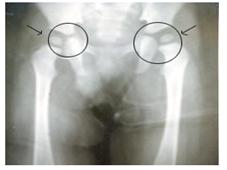
사진191.양쪽 정상 고관절
화살표로 표시된 ○내의 부위가 좌우 고관절이다
Copyright ⓒ 2011 John Sangwon Lee, MD., FAAP
| 선천성 고관절 탈구와 고관절 형성 이상의 원인 |
- 아직도 원인은 확실히 모른다.
- 분만 중 고관절에 상처가 생기면 고관절이 탈구될 수 있다.
- 선천성 만곡족 등 다리뼈나 발 뼈의 선천성 기형이 있을 때 그와 함께 이 병이 생길 수 있다.
- 태아의 자세, 즉 태위 이상이 있을 때 고관절이 탈구될 수 있다.
- 태아 때 호르몬의 영향으로도 생길 수 있다.
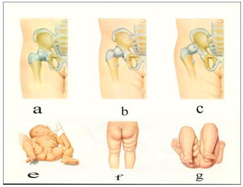
그림 192.정상 고관절과 고관절 탈구
a-정상 고관절, b-고관절 부전 탈구, c-고관절 완전 탈구, e-탈구된 우측 대퇴가 정상적으로 외전되지 않을 수 있다. f-양 대퇴부의 피부 주름살이 비대칭적일 수 있다. 우측에 비정상 주름살이 있다. g-양 대퇴의 길이에 차이가 생길 수 있다.
Used with permission from Clinical Educational Aid Ross Laboratories, Columbus, Ohio, USA과 소아가정간호백과
| 선천성 고관절 탈구와 고관절 형성 이상의 증상 징후 |
- 이 병은 유전으로 생길 수 있다.
- 때문에 어떤 가족에게는 이 병이 더 생길 가능성이 더 많다.
- 첫 번째로 태어나는 아기, 여아, 둔부 분만으로 태어난 아기에게 더 잘 생긴다.
- 고관절 형성 이상이나 고관절 부전 탈구는 100명의 신생아들 중 1명꼴로 생기고 고관절 탈구는 1000명의 신생아들 중 1.5명에게 생긴다.
- 갓 태어난 신생아나 그 후 영아의 한 쪽의 고관절에 선천성 고관절 탈구가 있으면 양 허벅다리의 주름살의 크기와 수가 다를 수 있고 양 다리의 길이에 차이가 날 수 있다.
- 탈구된 쪽 허벅다리를 양 옆으로 벌릴 때(그림 192-e) 탈구가 되지 않은 정상 허벅다리를 옆으로 벌릴 때보다 조금 덜 벌려진다.
- 기저귀를 채우기 위해서 양 허벅다리를 양 쪽 옆으로 벌릴 때 탈구된 쪽 허벅다리가 옆으로 정상적으로 잘 벌려지지 않을 수 있다.
- 탈구가 된 쪽 고관절을 손으로 만져서 진찰할 때 대퇴골의 두부가 고관절 고난절 낭속에서 탈구되어 있는 촉감을 느낄 수 있다.
- 탈구된 쪽 허벅다리에 생긴 피부 주름살의 수나 모양은 정상 허벅다리에 생긴 것들과 서로 다를 수 있다.
- 그러나 양쪽 고관절이 정상일 때도 양쪽 허벅다리의 주름살의 수나 모양이 서로 다를 수 있다.
- 이 병을 조기에 진단해서 적절히 치료해 주지 않으면 완전히 낫지 않고 일생동안 탈구된 상태로 산다.
- 아이가 서고 걷기 시작할 때부터 다리를 절 수 있다.
- 자라서 걷기 시작할 때 오리걸음같이 뒤뚱거린다.
- 이 병을 제때에 적절히 치료해도 완전히 치료되지 않아 일생 동안 절름거리는 불구자가 될 수 있다.
| 선천성 고관절 탈구와 고관절 형성 이상의 진단과 치료 |
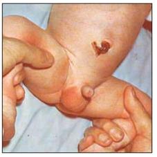
사진 193.선천성 고관절 탈구를 진단하기 위해 갓 태어난 신생아의 고관절을 통상적으로 진찰한다. 바른쪽 고관절 낭속에 있는 대퇴골 두부외 회전에 제한이 있다.
Used with permission from Mead Johnson Nutritional Division, 1972과 소아가정간호백과
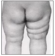
사진 194.선천성 고관절 탈구가 있으면 양 허벅다리의 주름살 수와 모양이 다를 수 있다. 그러나 신생아의 허벅다리 주름살 수와 모양이 다를 수 있다.
Used with permission from Mead Johnson Nutritional Division, 1972과 소아가정간호백과
- 병력, 증상 징후, 진찰소견 등을 종합해서 이 병이 의심되면 양쪽 고관절 초음파 검사 또는 X선 사진으로 진단한다.
- 처음 진단을 받을 때의 아기의 나이, 이 병의 정도와 종류 등에 따라 치료를 달리한다.
- 갓 태어나서 첫 돌까지 정기 건강검진을 받을 때 이 병이 있는지 기본적으로 진찰한다.
- 이 병이 의심되면 정형외과 전문의에 의뢰해서 정확한 진단을 받고 그에 따른 치료를 받는다.
- 정형외과에서 파브리크(Pavlik) 장치로 치료한다.
- 그 치료 효과는 상당히 좋다.
- 고관절에서 탈구된 대퇴골 근위 두부를 고관절 관절 낭속에 넣은 후 고관절을 깁스 해 고정시켜 치료하기도 한다.
- [부모도 반의사가 되어야 한다- 소아가정간호백과]-제 16권 소아청소년 정형외과 질환-척주측만증 진단과 치료 참조.
|
다음은 “뼈에서 딱딱 소리가 나요, 고관절 탈구”에 관한 인터넷 소아청소년 건강상담 질의응답의 예 입니다. |
Q&A. 뼈에서 딱딱 소리가 나요
Q.
2개월19일 된 남자 아이입니다. 어느 날인가 부터 관절에서 딱딱 소리가 나구요. 허벅지의 살 접히는 부분이 한쪽은 하나고 다른 쪽은 두개로 서로 다릅니다. 책에서 보니깐 고관절탈구가 그런 증상징후가 있다고 하는데 병원에 가야 하는지 가면 어떤 치료를 받는지 궁금해요.
좋은 답변 부탁드립니다.
A.
정미님
안녕하세요. 질문해 주셔서 감사합니다. 좋은 질문입니다.
자녀의 나이, 성별, 과거 병력, 가족 병력, 진찰소견, 임상검사 등의 정보를 많이 알수록 답변을 드리는데 도움이 됩니다. 주신 정보를 토대로 해서 답변을 드리겠습니다.
소아청소년과 전문의들은 아기의 고관절이 탈구되어 있나 알아보기 위해 태어난 이후부터 생후 6개월까지 정기 건강검진을 할 때마다 조심히 고관절을 특별히 진찰하는 것이 보통입니다.
그렇지만 진찰만 해서 고관절 탈구가 있나 없나를 확실히 진단하기가 쉽지 않습니다.
양쪽 허벅다리의 주름살의 크기와 개수의 차이 또는 다른 진찰 상 특이한 징후와 소견 등을 발견해 고관절 탈구를 의심해 볼 수는 있지만 고관절 초음파, X선 사진검사, 때로는 CT 스캔 검사 등으로 진단하기도 합니다.
그러나 영상과에서도 고관절 초음파 사진 검사나 고관절 CT 스캔 검사를 잘 판독하지 못해 오진하는 수 있습니다.
그 병에 경험이 많은 소아청소년 정형외과 전문의들이 이 병을 주로 진단치료를 하지만 그분들도 때로는 진단을 쉽게 붙이지 못할 수 있습니다.
태어 날 때 둔부선행 분만(둔위분만)으로 출생한 아기나 선천성 기형이 있는 아기 또는 다운 증후군이 있는 아기에게 이 기형이 좀 더 흔히 생길 수 있으나 사실은 아주 드문 선천성 병, 또는 후천성 병에 속합니다.
걱정이 되시면 정기 건강검진을 할 때 이 문제에 관해서 상담하시기 바랍니다.
[부모도 반의사가 되어야 한다– 소아가정간호백과]-제 16권 소아청소년 정형외과 질환–고관절 탈구, 고관절 형성 이상증을 참조하시기 바랍니다. 그리고 질문이 더 있으면 다시 연락해 주시기 바랍니다. 감사합니다. 이상원 드림
Congenital dislocation and dysplasia of the hip in newborn infants 선천성 고관절 탈구와 고관절 형성 이상
- The joint where the sciatic bone and the head of the femur are connected is called the hip joint.
- Some or all of the femoral head in the hip joint capsule may not develop naturally.
- A hip joint in which the head of the femur is not naturally formed in this way is called hip joint dysplasia.
- At this time, the head of the femur may come out of the hip joint capsule by birth. This disease is called congenital hip dislocation.
- Congenital hip dislocations can be crippled for life if not properly treated in time. Hip joint dislocation can occur when dysplasia occurs in one or more parts of the proximal head of the femur, or in several parts of the synovial membrane or other tissue surrounding the hip joint capsule, the sciatic joint where the head of the femur normally enters.
- When the hip joint is partially dislocated, it is called hip insufficiency dislocation, and when it is completely dislocated, it is called complete dislocation of the hip joint.
Picture 191. Both normal hip joints. The inner part indicated by the arrow is the left and right hip joints. Copyright ⓒ 2011 John Sangwon Lee, MD., FAAP
Causes of congenital hip dislocation and hip dysplasia
- Still not sure of the cause.
- If the hip joint is injured during delivery, the hip joint can be dislocated. Congenital clubfoot, congenital malformations of the back leg bones or foot bones can cause the disease along with it.
- The hip joint can be dislocated when there is an abnormal posture of the fetus. It can also be caused by hormonal effects in the fetus.
v
Figure 192. Normal hip and hip dislocation
The a-normal hip joint, b-hip insufficiency dislocation, c-complete hip dislocation, e-dislocation of the right thigh may not be abducted normally.
The skin folds of the f-lateral thighs may be asymmetric. There are abnormal wrinkles on the right side.
There may be a difference in the length of both g-legs. Used with permission from Clinical Educational Aid Ross Laboratories, Department of Columbus, Ohio, USA Department of Pediatric Home Nursing Symptoms of congenital hip dislocation and hip dysplasia
- This disease can be inherited.
- Because of this, some families are more likely to develop this disease.
- It is more prone to the first baby, girl, and baby born with a buttocks delivery.
- Hip dysplasia or hip dislocation occurs in 1 in 100 newborns, and hip dislocation occurs in 1.5 out of 1000 newborns.
- If a newborn or later infant has a congenital hip dislocation in one hip joint, the size and number of wrinkles on both thighs may differ, and the length of both legs may differ.
- When the dislocated thigh is spread to the sides (Fig. 192-e), it is slightly less open than when the undislocated normal thigh is spread to the side.
- When spreading both thighs to the sides to fill the diaper, the dislocated thigh may not open normally to the side.
- When examining the dislocated hip joint by touching it with your hand, you can feel the touch of the head of the femur being dislocated in the hip joint’s hip joint sac.
- The number or shape of skin folds on the dislocated thigh may differ from those on the normal thigh.
- However, even when both hip joints are normal, the number and shape of wrinkles on both thighs may be different. If this disease is not diagnosed early and treated properly, it will not completely heal and live in a dislocated state for life.
- Your child can limp from the moment your child starts to stand and walk. When I grow up and start walking, I stumble like a duck step.
- Even if the disease is treated properly and in time, it is not completely cured and can lead to a lifelong crippled person.
Diagnosis and treatment of congenital hip dislocation and hip dysplasia
Picture 193. In order to diagnose congenital hip dislocation, the hip joint of a newborn baby is usually examined.
There is a limitation of extra-headed rotation of the femur in the right hip sac. Used with permission from Mead Johnson Nutritional Division, Department 1972 Pediatric and Family Nursing Encyclopedia
Photo 194.
If there is a congenital hip dislocation, the number and shape of wrinkles on both thighs may be different.
However, the number and shape of the wrinkles on the thighs of the newborn can be different. Used with permission from Mead Johnson Nutritional Division, Department 1972 Pediatric and Family Nursing Encyclopedia
If the disease is suspected by taking the medical history, symptoms, signs, and examination findings together, it is diagnosed by ultrasound of both hips or X-rays.
Treatment differs depending on the age of the baby at the time of initial diagnosis and the severity and type of this disease.
When you receive a regular medical checkup from birth to the first birthday, you are basically checked for this disease.
If this disease is suspected, refer to an orthopedic specialist for accurate diagnosis and treatment accordingly.
In orthopedic surgery, it is treated with a Pavlik device.
The therapeutic effect is quite good.
In some cases, the head of the proximal femur dislocated from the hip joint is inserted into the hip joint capsule, and then the hip joint is cast and fixed. Visit www.drleepediatrics.com-Vol. 16 Pediatric and adolescent Orthopedic Diseases-See Diagnosis and Treatment of Spinal Scoliosis.
The following is an example of a question-and-answer on Internet pediatric and adolescent health counseling about “cracking in the bones, hip dislocation”.
Q&A.
There is a crackling sound in the bones
Q.
This is a boy who is 2 months and 19 days old. From one day, the joints crackle. The folds of the thigh are different, one on one side and two on the other. The book says that hip dislocation has such symptoms, but I am curious to see if I need to go to the hospital or what kind of treatment will I get if I go there. Please give me a good answer.
A.
Jeongmi
Good morning. Thanks for asking.
That’s a good question.
The more information you know about your child’s age, gender, past medical history, family medical history, medical examination findings, and clinical examination, the more helpful it is to give you an answer.
We will respond based on the information you provided.
It is common for pediatricians and adolescents to carefully examine the hip joint every time they perform regular health check-ups from birth to 6 months of age to see if the baby’s hip joint is dislocated.
However, it is not easy to diagnose for sure whether there is a dislocation of the hip joint just by examining it.
Although it is possible to suspect hip dislocation by discovering the difference in the size and number of wrinkles on both thighs, or by finding unusual signs and findings on other examinations, it is also diagnosed by ultrasound of the hip joint, X-ray photographic examination, and sometimes CT scan.
However, the imaging department may also misdiagnose the hip joint ultrasound photo test or hip joint CT scan test because it is not well-read.
Pediatric and adolescent orthopedic specialists who are experienced with the disease mainly diagnose and treat the disease, but sometimes they may not be able to attach a diagnosis easily.
This anomaly may be more common in babies born at birth as a result of pre-granular delivery (bull delivery), congenital anomalies, or babies with Down syndrome, but it is in fact a very rare congenital or acquired disease. If you are concerned, please consult with us about this matter during regular health check-ups.
Visit www.drleepediatrics.com]-Volume 16 Children and Adolescents Orthopedic Diseases-Hip Dislocation, Hip dysplasia, please refer. And if you have more questions, please contact us again. Thank you. Lee Sang-won dream
출처 및 참조 문헌
- NelsonTextbook of Pediatrics 22ND Ed
- The Harriet Lane Handbook 22ND Ed
- Growth and development of the children
- Red Book 32nd Ed 2021-2024
- Neonatal Resuscitation, American Academy Pediatrics
- www.drleepediatrics.com 제1권 소아청소년 응급 의료
- www.drleepediatrics.com 제2권 소아청소년 예방
- www.drleepediatrics.com 제3권 소아청소년 성장 발육 육아
- www.drleepediatrics.com 제4권 모유,모유수유, 이유
- www.drleepediatrics.com 제5권 인공영양, 우유, 이유식, 비타민, 미네랄, 단백질, 탄수화물, 지방
- www.drleepediatrics.com 제6권 신생아 성장 발육 육아 질병
- www.drleepediatrics.com제7권 소아청소년 감염병
- www.drleepediatrics.com제8권 소아청소년 호흡기 질환
- www.drleepediatrics.com제9권 소아청소년 소화기 질환
- www.drleepediatrics.com제10권. 소아청소년 신장 비뇨 생식기 질환
- www.drleepediatrics.com제11권. 소아청소년 심장 혈관계 질환
- www.drleepediatrics.com제12권. 소아청소년 신경 정신 질환, 행동 수면 문제
- www.drleepediatrics.com제13권. 소아청소년 혈액, 림프, 종양 질환
- www.drleepediatrics.com제14권. 소아청소년 내분비, 유전, 염색체, 대사, 희귀병
- www.drleepediatrics.com제15권. 소아청소년 알레르기, 자가 면역질환
- www.drleepediatrics.com제16권. 소아청소년 정형외과 질환
- www.drleepediatrics.com제17권. 소아청소년 피부 질환
- www.drleepediatrics.com제18권. 소아청소년 이비인후(귀 코 인두 후두) 질환
- www.drleepediatrics.com제19권. 소아청소년 안과 (눈)질환
- www.drleepediatrics.com 제20권 소아청소년 이 (치아)질환
- www.drleepediatrics.com 제21권 소아청소년 가정 학교 간호
- www.drleepediatrics.com 제22권 아들 딸 이렇게 사랑해 키우세요
- www.drleepediatrics.com 제23권 사춘기 아이들의 성장 발육 질병
- www.drleepediatrics.com 제24권 소아청소년 성교육
- www.drleepediatrics.com 제25권 임신, 분만, 출산, 신생아 돌보기
- Red book 29th-31st edition 2021
- Nelson Text Book of Pediatrics 19th- 21st Edition
- The Johns Hopkins Hospital, The Harriet Lane Handbook, 22nd edition
- 응급환자관리 정담미디어
- Pediatric Nutritional Handbook American Academy of Pediatrics
- 소아가정간호백과–부모도 반의사가 되어야 한다, 이상원 저
- The pregnancy Bible. By Joan stone, MD. Keith Eddleman, MD
- Neonatology Jeffrey J. Pomerance, C. Joan Richardson
- Preparation for Birth. Beverly Savage and Dianna Smith
- 임신에서 신생아 돌보기까지. 이상원
- Breastfeeding. by Ruth Lawrence and Robert Lawrence
- Sources and references on Growth, Development, Cares, and Diseases of Newborn Infants
- Emergency Medical Service for Children, By Ross Lab. May 1989. p.10
- Emergency care, Harvey Grant and Robert Murray
- Emergency Care Transportation of Sick and Injured American Academy of Orthopaedic Surgeons
- Emergency Pediatrics A Guide to Ambulatory Care, Roger M. Barkin, Peter Rosen
- Quick Reference To Pediatric Emergencies, Delmer J. Pascoe, M.D., Moses Grossman, M.D. with 26 contributors
- Neonatal resuscitation Ameican academy of pediatrics
- Pediatric Nutritional Handbook American Academy of Pediatrics
- Pediatric Resuscitation Pediatric Clinics of North America, Stephen M. Schexnayder, M.D.
-
Pediatric Critical Care, Pediatric Clinics of North America, James P. Orlowski, M.D.
-
Preparation for Birth. Beverly Savage and Dianna Smith
-
Infectious disease of children, Saul Krugman, Samuel L Katz, Ann A.
- 제4권 모유, 모유수유, 이유 참조문헌 및 출처
- 제5권 인공영양, 우유, 이유, 비타민, 단백질, 지방 탄수 화물 참조문헌 및 출처
- 제6권 신생아 성장발육 양호 질병 참조문헌 및 출처
- 소아과학 대한교과서
Copyright ⓒ 2014 John Sangwon Lee, MD, FAAP
“부모도 반의사가 되어야 한다”-내용은 여러분들의 의사로부터 얻은 정보와 진료를 대신할 수 없습니다.
“The information contained in this publication should not be used as a substitute for the medical care and advice of your doctor. There may be variations in treatment that your doctor may recommend based on individual facts and circumstances.
“Parental education is the best medicine.